Hepatobiliary and Pancreatic Unit

Anatomy of the bile ducts
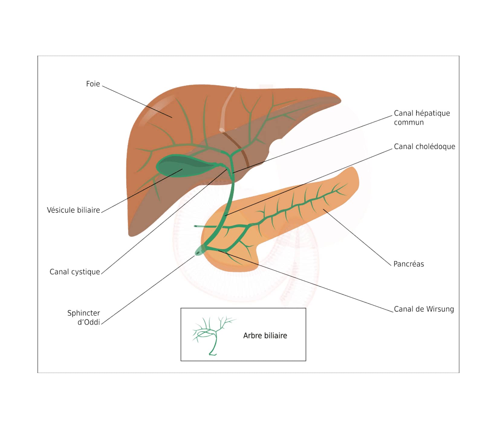
The intrahepatic bile ducts are formed by a set of small ducts and unite into two right and left hepatic ducts (ducts leaving the liver). These ducts form a common hepatic duct which becomes extrahepatic.
The extrahepatic bile ducts include:
- a) The common bile duct which is made up of the union of the two right and left hepatic ducts thus forming the common hepatic duct. This duct joins the cystic duct which is itself connected to the gallbladder, it then becomes the bile duct, which descends behind the pancreas and ends in the second portion of the duodenum at the level of the ampulla of Vater and the sphincter of Oddi.
- b) The gallbladder connected by the cystic duct to the main duct is a reservoir of bile which is discharged into the duodenum at the time of feeding to mix with the bolus and allow digestion.
Gallbladder cancer
The gallbladder is a very small hollow organ located under the liver. it is a reservoir of bile and helps us maintain a healthy digestion.
Gallbladder cancer is one of the most common cancers affecting the bile ducts. If this pathology is diagnosed early enough, the cancer can be cured by performing a resection of the gallbladder. It is twice as common in women as in men.

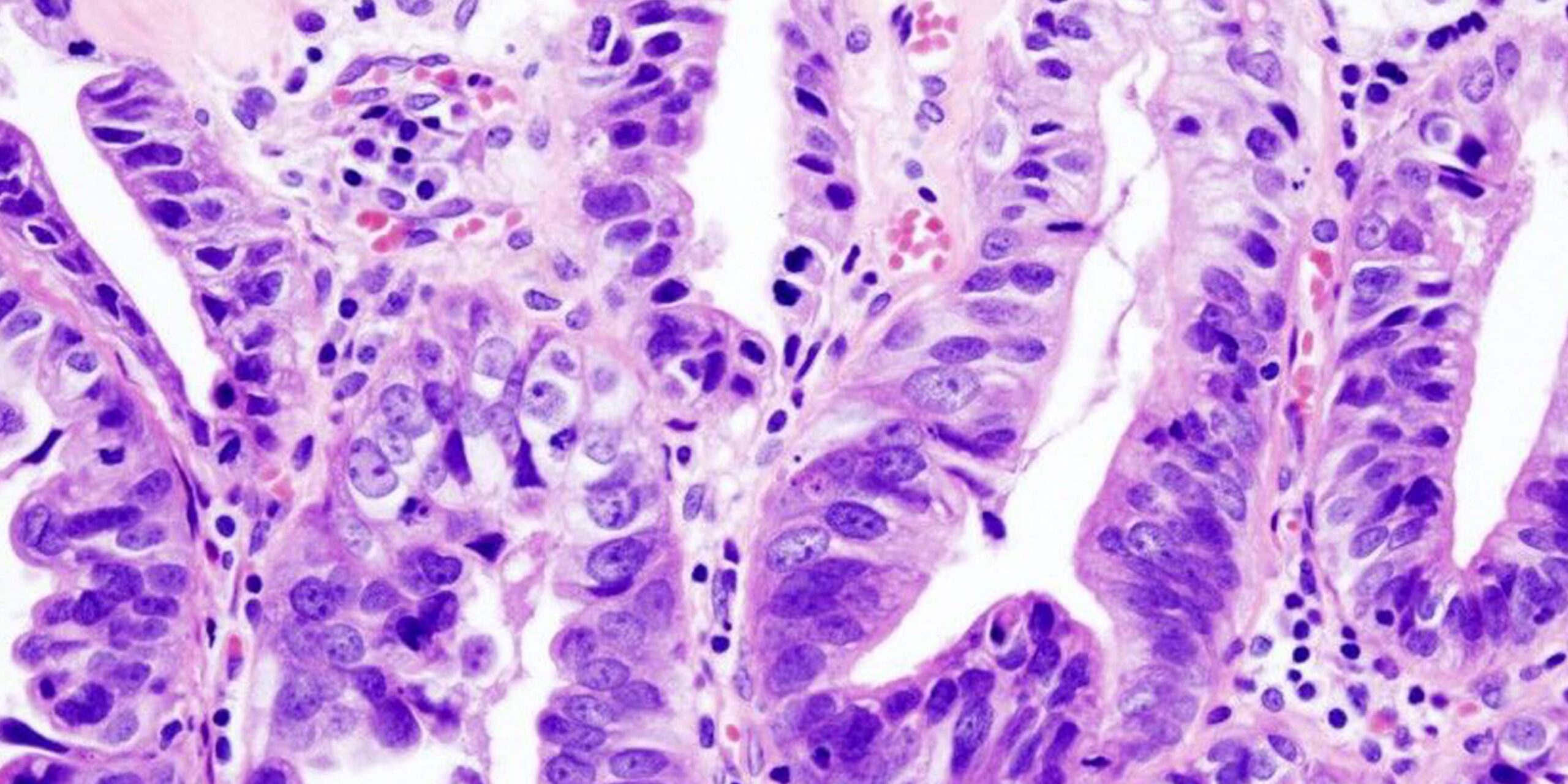
Histopathology of gallbladder adenocarcinoma, Wikimedia Commons
Risk factors
Several factors can promote the appearance of this cancer:
- obesity
- cholelithiasis
- chronic inflammation
- age
- anatomical abnormalities between the common bile duct and the main pancreatic duct
Symptoms
Initially, the disease is asymptomatic; the first symptoms mimic inflammation of the gallbladder due to gallstones.
The following signs may appear:
- abdominal pain,
- bloating of the abdomen,
- nausea, vomiting and dizziness,
- fever which may be accompanied by jaundice,
- weight loss,
- loss of appetite
- bad breath,
- digestion problems and gas.
Diagnosis and management
Gallbladder cancer is often discovered incidentally after laparoscopic cholecystectomy for gallbladder lithiasis.
Particular attention must be taken to verify that the symptoms of cancer are indeed present. The medical history and physical examination of the patient are also very important in diagnosing the disease.
A liver MRI with Bili-IRM is most often necessary to accurately assess the extent of the lesion.
An extension assessment with the dosage of tumor markers (Ca 19-9), a liver assessment and a thoraco-abdominal scan must be carried out to assess the disease.
Treatment – Post-operative follow-up
The most common and effective treatment for cure is surgical removal of the gallbladder (cholecystectomy) along with part of the liver and lymph nodes. Chemotherapy may be offered before surgery to reduce the size of the tumor.
If surgical treatment is impossible due to an advanced stage of the disease, endoscopic biliary prosthesis placement can reduce jaundice and relieve vomiting.
After complete resection, additional so-called “adjuvant” chemotherapy may be offered to reduce the risk of recurrence.
Useful information
Download guide: Liver cancer treatments
by National Cancer Institute, e-cancer.fr
Cancer Info Line
Support throughout your care pathway
Quality patient care is an essential objective, and the patient is at the heart of our concerns.


